
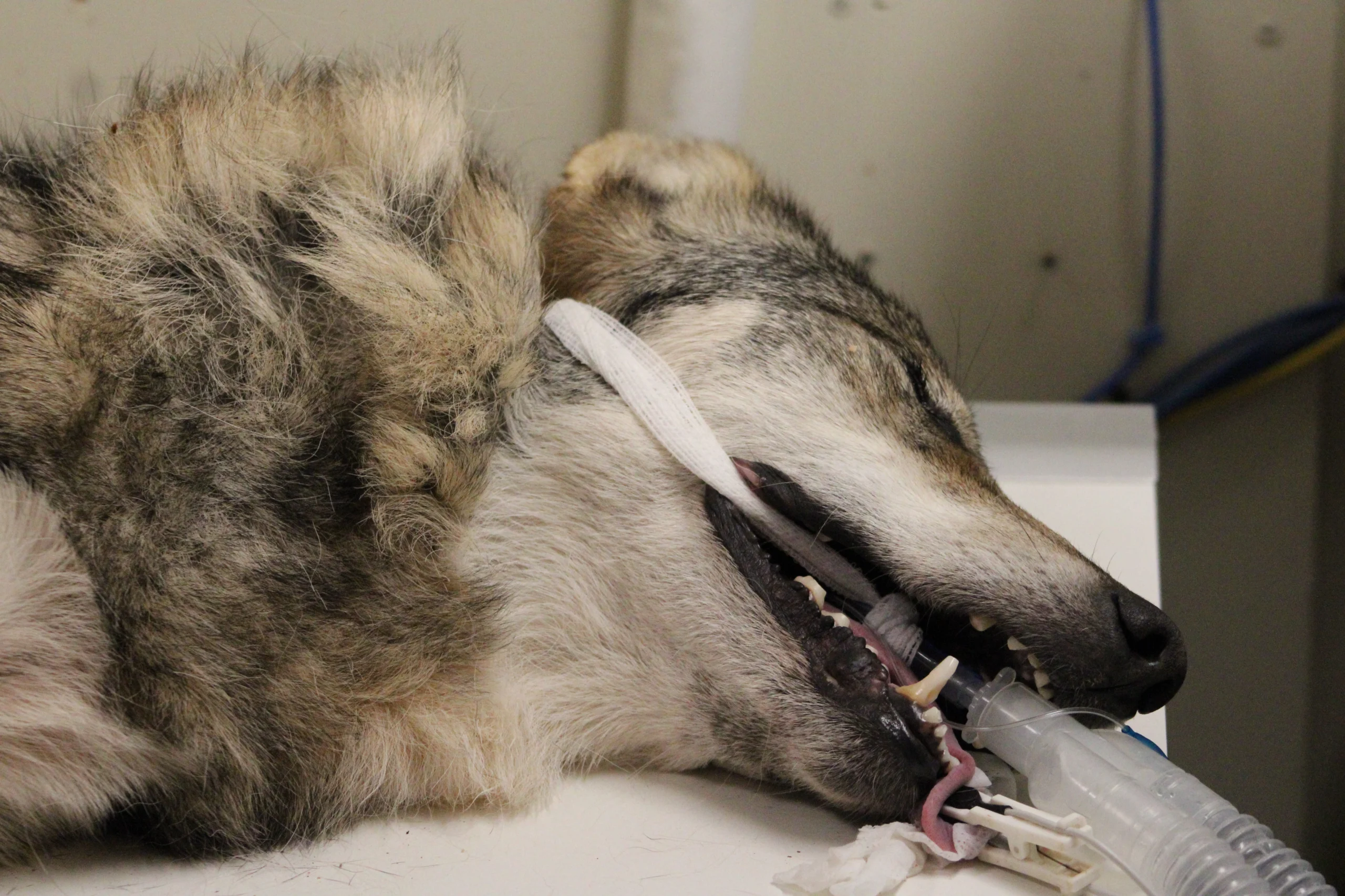
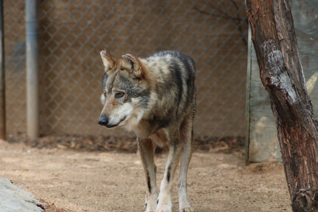
Angus, the Mexican gray wolf, came into the clinic on October 6th for his annual exam. Part of his exam included checking his teeth and mouth to make sure everything was healthy, especially because Mexican gray wolves are prone to oral and nasal cancers. Although the reason for this is unknown, a genetic link is suspected.
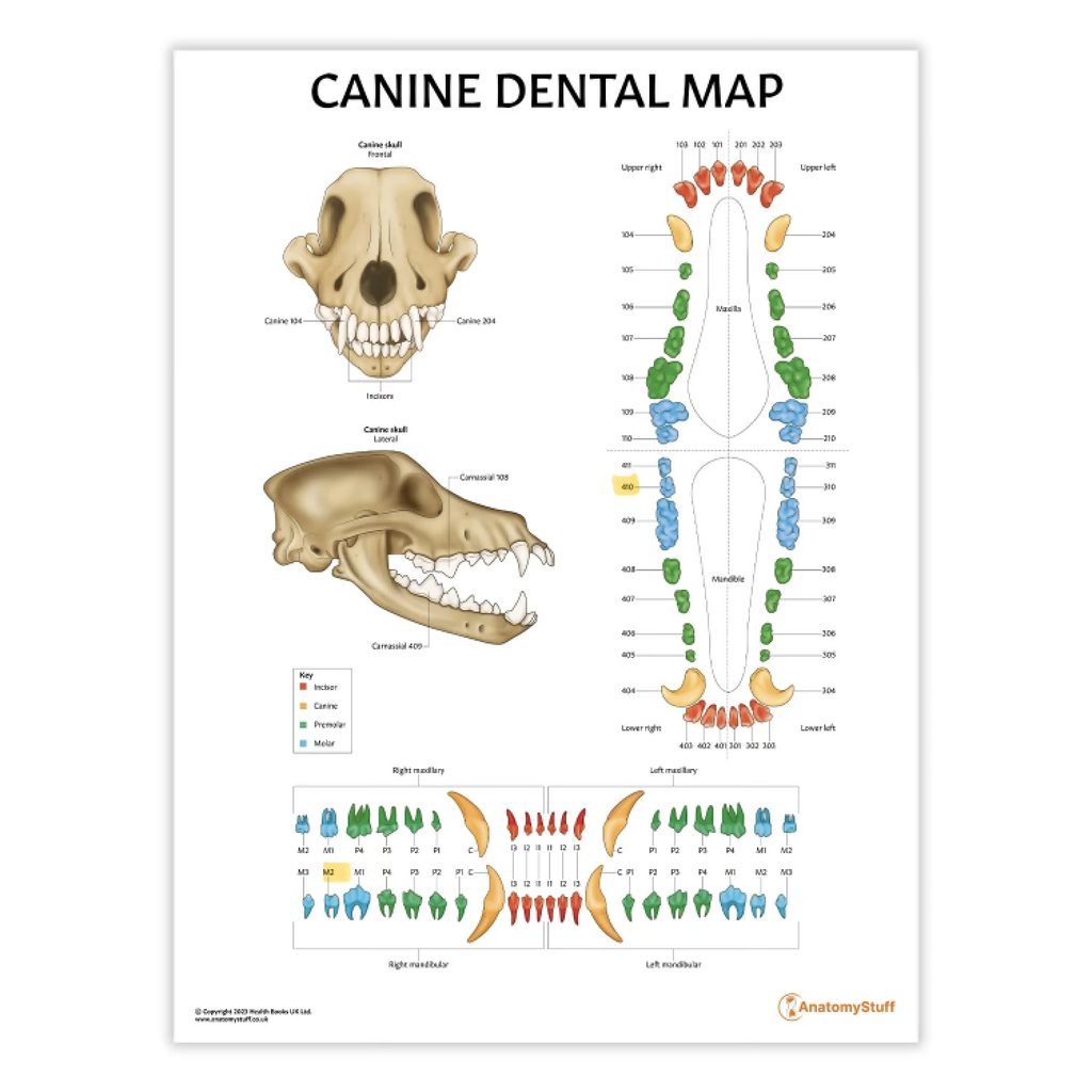
During his oral exam, a dental probe was used to check the gumline around each tooth for signs of dental disease. When the veterinarian, Dr. Woodburn, examined one of Angus’s teeth, she noticed that the probe sank into the gumline and caused bleeding. The affected tooth was one of his lower right molars, referred to as tooth “410” in veterinary dentistry.
Fun fact: Each tooth has its own number for medical reference, just as in human dentistry!
Because this molar plays an important role in chewing, the dental disease was likely causing Angus significant discomfort. To prevent further pain, we determined that the tooth needed to be extracted. Below are just a few of the factors our team considers. Every animal and every rehabilitation case is unique, so the care leading up to release is individualized to meet that animal’s needs. For some, that may mean spending additional time in our clinic or making several trips back for veterinary care to ensure an injury has healed properly. Others may benefit from being paired with another animal of the same species and of a similar age and size as they develop and practice natural behaviors. Rehabilitation takes time, observation, and adjustments along the way.
Before the extraction could begin, some preparation was needed. Just like when humans visit the dentist, plaque must be removed before a tooth is extracted to reduce the risk of introducing bacteria into the extraction site and causing an infection. While Dr. Woodburn prepared her extraction tools, our Clinic Manager, Dani, scaled and polished all of Angus’s teeth to reduce the bacteria in his mouth. This process is much like a routine dental cleaning at your own dentist’s office.
Once the cleaning was complete, Dr. Woodburn began the extraction. This is a slow, delicate process because improper technique can damage the jawbone or surrounding teeth. First, she numbed the area with a lidocaine injection and carefully cut the gum tissue around the affected tooth to create what is called a gingival flap. This allows the gums to be sutured closed after the tooth is removed.
Next, she used a dental drill to divide the tooth down the middle. Because this molar has two roots, separating it into two sections makes the extraction easier and reduces stress on the jawbone. She then carefully loosened each section from the surrounding bone and removed it! Dental X-rays were taken afterward to confirm that the entire tooth had been extracted. Once everything looked good, Dr. Woodburn sutured the gum tissue closed, and Angus’s dental procedure was complete!
The entire process, including the rest of his annual exam, took about three hours. Medical staff closely monitored Angus throughout the procedure, and thankfully, he did very well under anesthesia. Given his advanced age, we decided to complete the extraction that day rather than postpone it. Anesthesia can carry greater risks for older animals, and since Angus was tolerating it well, we wanted to avoid putting him through another anesthetic procedure on a separate day.
After recovering from anesthesia, Angus was able to return to his enclosure. His post-operative care included a few days of pain medication and a soft diet for 10–14 days. Our Animal Care team provided soft meats and monitored his eating habits throughout his recovery.
Fortunately, Angus healed well, and there have been no complications! His experience is a great example of the careful monitoring and individualized medical care that help us support the health and well-being of the wildlife in our care!
October, 2026